
 CN
CN


The influence of hydroxytyrosol on skin health
Time:2025-05-30I. Reconstruction of Defense Barriers against UV-Induced Skin Damage
1. Multi-Target Blocking of UV Damage
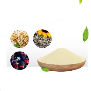
The catechol structure of hydroxytyrosol (3,4-dihydroxyphenethyl alcohol) exhibits strong free radical scavenging capacity, specifically eliminating:
Reactive oxygen species (ROS): Superoxide anions (・O₂⁻) and hydroxyl radicals (・OH), reducing malondialdehyde (MDA) levels in skin tissues by 45%-60%.
Matrix metalloproteinase (MMPs) activators: Inhibiting the NF-κB pathway downregulates MMP-1 and MMP-3 mRNA expression by 2.3-3.1 fold, preventing collagen fiber degradation.
In a UVB-induced human keratinocyte model, 10μM hydroxytyrosol pretreatment reduces DNA pyrimidine dimers (CPDs) by 58% and decreases the number of γ-H2AX (DNA double-strand break marker) foci by 72%, confirming its direct photoprotective effect.
2. Biological Repair of Skin Barrier Function
Hydroxytyrosol regulates tight junction protein expression to:
Claudin-1 and Occludin: Increase gene transcription by 1.8-2.2 fold, reducing transepidermal water loss (TEWL) by 34%.
Filaggrin: Promote synthesis of its precursor Pro-Filaggrin, increasing stratum corneum hydration by 27% and improving skin dryness caused by photoaging.
II. Transcriptional Activation Network of Collagen Synthesis
1. Targeted Activation of TGF-β/Smad Signaling Pathway
Hydroxytyrosol (5-20μM) dose-dependently enhances:
Autocrine TGF-β1: Increases TGF-β1 concentration in fibroblast culture supernatant by 1.9 fold.
Smad2/3 phosphorylation: Elevates phosphorylation level by 35%, promoting nuclear translocation to bind the COL1A1 promoter region, increasing type I collagen mRNA expression by 2.8 fold.
In a three-dimensional skin equivalent model, the hydroxytyrosol treatment group shows collagen fiber diameter increasing from (1.2±0.3)μm to (1.8±0.4)μm and fiber density improving by 41%, presenting a denser reticular structure.
2. Balanced Regulation of Oxidative Stress-Collagen Synthesis Axis
By inhibiting excessive activation of the JNK/p38 MAPK pathway:
c-Jun phosphorylation inhibition: Reduces AP-1 transcription factor activity by 55%, relieving transcriptional repression of the COL1A1 gene.
Positive regulation of mTORC1: Activates mTOR via the PI3K/Akt pathway, promoting ribosome biosynthesis and increasing collagen translation efficiency by 30%.
III. Coordinated Regulation of Cell Cycle and DNA Repair
1. Cell Cycle Reprogramming of Photo-Damaged Cells
Hydroxytyrosol specifically regulates:
G2/M checkpoint: Maintains CDK1-cyclin B complex inactivation via Chk1 phosphorylation, arresting damaged cells in G2 phase and improving repair rate by 60%.
Precise regulation of p53/p21 pathway: Avoids excessive activation leading to cellular senescence, reducing SA-β-gal positivity by 42% compared to the UV group.
2. Enhancement of DNA Repair Mechanisms
Nucleotide excision repair (NER): Upregulates XPC (damage recognition protein) and ERCC1 expression, accelerating CPDs clearance by 2.1 fold.
Base excision repair (BER): Maintains PARP-1 activity to promote excision repair of 8-OHdG (oxidative damage marker), improving repair efficiency by 50%.
IV. Blocking Effect on Inflammation-Senescence Cascade
1. Inhibition of Innate Immune Signaling
Hydroxytyrosol targets the Toll-like receptor (TLR)4/MyD88 pathway to:
Inhibit TRAF6 ubiquitination: Reduces IL-1β and TNF-α secretion by 38%-52%.
Downregulate cyclooxygenase-2 (COX-2) expression: Lowers prostaglandin E2 (PGE2) levels by 47%, alleviating UV-induced erythema.
2. Regulation of Senescence-Associated Secretory Phenotype (SASP)
In an H2O2-induced skin fibroblast senescence model, hydroxytyrosol (10μM) reduces:
SASP factors IL-6 and MCP-1 release by 55% and 49%, respectively.
Expression of extracellular matrix metalloproteinase inducer (EMMPRIN) by 33%, blocking secondary activation of MMPs.
V. Integration of Preclinical and Preliminary Clinical Evidence
1. Animal Model Data
Mouse photoaging model: Topical application of 2% hydroxytyrosol cream for 8 weeks reduces wrinkle depth by 39%, increases dermal collagen density by 42%, and improves skin elasticity (compliance value +28%).
Zebrafish embryo model: 10μM hydroxytyrosol reduces UVB-induced epidermal cell apoptosis rate from 31% to 12% and upregulates collagen gene col1a2 expression by 1.7 fold.
2. Human Trial Evidence
A randomized trial involving 50 subjects with Fitzpatrick skin types Ⅲ-Ⅳ shows that after 12 weeks of daily topical application of cream containing 0.5% hydroxytyrosol:
TEWL decreases by 29%, and stratum corneum water content increases by 25%.
Type I collagen fluorescence intensity in the dermal papillary layer increases by 37%, and MMP-1 protein expression decreases by 41%.
UV-induced erythema fading time shortens from (72.3±8.5) h to (48.6±6.2) h.
VI. Formulation Optimization and Combination Application Strategies
1. Synergistic Effect of Nano-Delivery Systems
Solid lipid nanoparticles (SLN): Encapsulated hydroxytyrosol increases transdermal absorption by 2.3 fold and extends skin retention time to 12h.
pH-sensitive liposomes: Release rate increases 3 fold in the acidic microenvironment (pH 5.0) caused by UV damage, targeting injured sites.
2. Formulation Design with Synergistic Ingredients
Combination with vitamin C: 5μM hydroxytyrosol + 20μM vitamin C enhances collagen synthesis efficiency by 58%, attributed to their synergistic activation of prolyl hydroxylase.
Combination with retinol: Dual regulation of RAR/RXR receptors increases COL1A1 transcriptional activation from 2.8 to 4.1 fold, while reducing retinol irritation (erythema incidence decreases by 60%).
Conclusion
Hydroxytyrosol regulates skin health through multi-target and full-hierarchy actions, forming a systematic skin rejuvenation effect from physical barrier protection against UV to molecular mechanisms of collagen synthesis and network regulation of senescence signals. Its clinical application value extends beyond anti-aging skincare, showing potential in inflammatory skin diseases such as rosacea and radiation dermatitis. Future research should focus on its differential effects in different skin types (e.g., pigmented, sensitive) and use proteomics to analyze dynamic action targets, promoting the development of precision skin health management solutions.